|
|
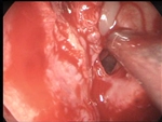
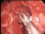
| 1. Starting sphenoethmoidectomy on the left. |
2. Removal of polyps and cells from the middle nasal meatus. |
|
|
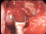
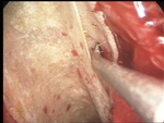
| 3. Removal of a maxillary sinus cyst. |
4. Entering the posterior ethmoids. |
|
|
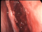
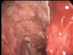
| 5. Locating the superior nasal concha. |
6. Posterior to anterior ethmoidectomy. |
|
|
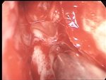
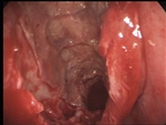
| 7. Correction of septal deviation. |
8. Right ethmoidectomy. |
|
|
| 9. The right superior nasal concha. |
10. Entering agger nasi. |
|
|
| 11. Starting the process of flattening the fovea ethmoidalis and lamina papyracea. |
12. Removal of the polypoid mucosa with Weil Blakesley forceps…. |
|
|
| 13. … or with a Cottle elevator. |
14. Trying to remove mucosa around the anterior ethmoid artery. |
|
|
| 15. Removal of bony septa with a diamond burr. |
16. Flattening the lamina papyracea. |
|
|
| 17. The removal of the mucosa probably implies the removal of the periosteum also. |
18. A bleeding point behind the posterior ethmoid artery. |
|
|
| 19. General view of the left nasal cavity. |
20. Right nasal cavity. Starting flattening process. |
|
|
| 21. Elevation of the mucosa with a curette. |
22. Removal of bony septa with a diamond burr. |
|
|
| 23. Tissue removal with a Weil Blakesley forceps |
24. Flattening the lamina papyracea. |
|
|
| 25. A bleeding point in front of the sphenoid sinus. |
26. Haemostasis with surgicel. |
|
|
| 27. Continue smoothing the bone projections. |
28. Covering the cavity until the graft insertion. |
|
|
| 29. Suture to fixate the middle turbinates. |
30. Passing the suture through the right concha. |
|
|
| 31. Tying the suture in the left nasal cavity. |
32. Preparing the area to take the graft. |
|
|
| 33. Taking the graft. |
34. Split-thickness skin grafts. |
|
|
| 35. Preparation to insert the graft into the left nasal cavity. |
36. Removing tamponade and surgicel. |
|
|
| 37. Insertion of the graft with the paper. |
38. Trying to apply the graft into the surgical cavity and …. |
|
|
| 39. … detach it from the paper. |
40. The process is almost complete. |
|
|
| 41. Removing the paper. |
42. Final view. No packing is needed. |
|
|
| 43. Removing the packing from the right nasal cavity. |
44. Insertion of the graft with the paper. |
|
|
| 45. Pressing the graft for application on the surgical cavity. |
46. Detaching the paper (middle concha). |
|
|
| 47. Detaching the paper (lamina papyracea). |
48. Detaching the paper (sphenoid sinus). |
|
|
| 49. Almost complete detachment of the paper. |
50. Removing air between the graft and the bone. |
|
|
| 51. Final view. No packing is needed. |
52. The skin graft one month after the operation. |
|


