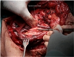
Functional Neck Dissection
Modified Radical Neck Dissection type III
| 1. The incision. A block of metastatic nodes is noted. | 2. Elevating the anterior flap. |
| 3. The relation of the marginal mandibular branch of the facial nerve to the facial vessels. | 4. Completion of the dissection of the submental triangle. Beginning dissecting the submandibular area. |
| 5. Identification of the hypoglossal nerve medially to the digastric muscle. | 6. Completing the dissection of the submandibular triangle. |
| 7. Starting dissection along the jugular chain. The specimen of the submandibular triangle has been removed. | 8. Descending hypoglossi and hypoglossal nerve. |
| 9. Ligation of the common facial vein. | 10. Identification of the omohyoid muscle. |
| 11. Completion of dissection to the level of the omohyoid muscle. We can see the inferior root anastomosing with the descending hypoglossi, and the superior thyroid artery. | 12. The same step of the operation but with an anterior and medial rotation of the specimen. |
| 13. Continuing dissecting the specimen of the anterior neck triangle free towards the midline, the vagus nerve is revealed. | 14. View after removal of the specimen. The ansa cervicalis is seen. |
| 15. Elevating the posterior cutaneous flap. The trapezius muscle and the accessory nerve are seen. | 16. While elevating the flap more cutaneous and muscular branches of the cervical plexus are revealed and must be distinguished from the accessory nerve. |
| 17. At the posterior triangle, the accessory nerve is situated about one centimeter above and parallel to the great auricular nerve. | 18. Furthermore, the accessory is situated at a higher level comparing to the branches of the cervical plexus. |
| 19. Finally, the accessory nerve enters medially to the trapezius muscle comparing to the superficial cutaneous branches of the cervical plexus. | 20. Mobilizing the accessory nerve. |
| 21. Ligating the suprascapular artery. | 22. Elevating the fibrous-fatty tissue from the supraclavicular fossa. |
| 23. By continuing elevating the specimen, the phrenic nerve and the brachial plexus are revealed. | 24. Elevating the specimen from the prevertebral muscles. |
| 25. Continuing elevating the specimen from the phrenic nerve. | 26. While elevating the fibrous-fatty tissue, two or three branches of the cervical plexus are divided. |
| 27. One step before the completion of the dissection. | 28. The surgical field after the removal of the specimen. |


